Table of Contents :
- Introduction
- The Heart’s Vital Role
- The Complexity of Heart Structure
- Anatomy of the Heart
- Chambers of the Heart
- Valves: Gatekeepers of Blood Flow
- Blood Vessels: The Circulatory Network
- Function of the Heart
- The Pumping Action
- Oxygen-Rich and Oxygen-Poor Blood
- Heartbeat: Systole and Diastole
- The Circulatory System
- Arteries, Veins, and Capillaries
- Pulmonary Circulation: To the Lungs and Back
- Systemic Circulation: Nourishing the Body
- Electrical System of the Heart
- SA Node: The Natural Pacemaker
- AV Node: Gatekeeper of Ventricular Contractions
- Impulse Conduction: Creating the Heartbeat
- Heart Sounds: Lub-Dub Rhythm
- S1 Sound: Closing of AV Valves
- S2 Sound: Closing of Semilunar Valves
- Listening to Heart Sounds
- Cardiac Cycle
- Atria Contraction: Filling the Ventricles
- Ventricular Contraction: Pumping Blood Out
- Relaxation: Preparing for the Next Cycle
- Heart Health and Disease
- Common Heart Conditions
- Risk Factors for Heart Disease
- Maintaining Heart Health
- Cardiovascular Research and Innovations
- Advancements in Cardiac Surgery
- Heart Transplants and Artificial Hearts
- Cutting-Edge Imaging Techniques
- Lifestyle and Heart Health
- Diet and Nutrition
- Exercise and Physical Activity
- Stress Management
- Conclusion
- The Heart’s Remarkable Journey
- Nurturing Your Heart for a Healthy Life
Introduction:
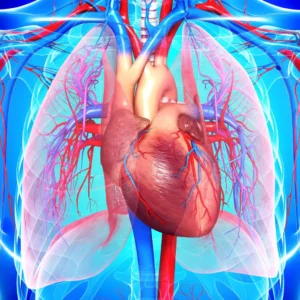
The human heart is a remarkable organ that plays a vital role in sustaining life. It serves as the central component of the cardiovascular system, responsible for pumping blood throughout the body. This intricate pump works tirelessly to ensure that oxygen, nutrients, hormones, and immune cells are efficiently distributed to various tissues and organs. The heart’s rhythmic contractions are essential for maintaining circulation, making it an indispensable organ for overall health and well-being.
The Heart’s Vital Role:
- Cardiovascular Centerpiece: The heart stands at the core of the body’s cardiovascular system, a complex network of blood vessels responsible for transporting essential substances.
- Circulation of Blood: Through its coordinated contractions, the heart propels blood into the circulatory pathways, allowing oxygen and nutrients to reach every corner of the body.
- Oxygen Delivery: The heart’s pumping action ensures that oxygen-enriched blood is delivered to tissues and organs, enabling their proper function.
- Waste Removal: As blood flows through the heart, waste products like carbon dioxide are collected for elimination from the body.
The Complexity of Heart Structure:
- Four-Chambered Design: The heart comprises four chambers: two atria (upper chambers) and two ventricles (lower chambers), each contributing to the heart’s efficient functioning.
- Atria Function: The atria receive blood from various parts of the body and lungs. The right atrium receives deoxygenated blood from the body, while the left atrium receives oxygenated blood from the lungs.
- Ventricles Function: The ventricles are responsible for pumping blood out of the heart. The right ventricle pumps deoxygenated blood to the lungs for oxygenation, and the left ventricle pumps oxygen-rich blood to the body.
- Valves and Pathways: The heart is equipped with valves that regulate the flow of blood between chambers and prevent backflow. These valves ensure unidirectional circulation.
The human heart’s importance goes beyond being a mere muscle; it’s the lifeline that sustains us. Its role in maintaining circulation, delivering oxygen and nutrients, and eliminating waste is unparalleled. Understanding the complexity of the heart’s structure lays the foundation for comprehending its incredible function, which is essential for our survival and well-being.
Anatomy of the Heart:

Chambers of the Heart:
- Atria: The atria act as receiving chambers. They collect blood returning from the body (right atrium) and oxygenated blood from the lungs (left atrium).
- Ventricles: The ventricles are responsible for pumping blood. The right ventricle pumps deoxygenated blood to the lungs, while the left ventricle propels oxygen-rich blood to the body.
Valves: Gatekeepers of Blood Flow:
- Tricuspid Valve: This valve prevents backflow of blood from the right ventricle to the right atrium.
- Pulmonary Valve: The pulmonary valve prevents blood from flowing back into the right ventricle after it has been pumped to the lungs.
- Mitral Valve: The mitral valve prevents blood from flowing back into the left atrium from the left ventricle.
- Aortic Valve: This valve prevents backflow of blood from the aorta into the left ventricle.
Blood Vessels: The Circulatory Network:
- Arteries: Arteries carry oxygenated blood away from the heart. The aorta, the largest artery, branches into arteries that deliver blood to various body regions.
- Veins: Veins transport deoxygenated blood back to the heart. The superior and inferior vena cava are major veins that return blood to the right atrium.
- Capillaries: Capillaries are where exchange occurs. Oxygen and nutrients diffuse from capillaries into tissues, while waste products move from tissues into capillaries.
Fascinating Facts:
- The heart pumps about 2,000 gallons (7,570 liters) of blood daily, which is enough to fill more than 60 tanker trucks.
- The human heart can create enough pressure to squirt blood 30 feet (9 meters) in the air.
- The “lub-dub” sound of a heartbeat is caused by the opening and closing of heart valves.
- Your heart can keep beating even when separated from the body due to its intrinsic electrical activity.
- Blood vessels in your body, if stretched end to end, could circle the Earth more than twice.
Understanding the intricacies of the heart’s anatomy, its various chambers, valves, and the extensive network of blood vessels showcases the extraordinary design of this essential organ. It tirelessly pumps blood, delivering oxygen and nutrients to every cell and tissue, ensuring the body’s survival.
Function of the Heart:

- The Pumping Action:
- The heart serves as a muscular pump, propelling blood throughout the body to deliver oxygen and nutrients to cells and remove waste products.
- The heart achieves this function through rhythmic contractions and relaxations.
- Oxygen-Rich and Oxygen-Poor Blood:
- Pulmonary Circulation: Deoxygenated blood returns to the right atrium from the body, then enters the right ventricle. This blood is pumped to the lungs via the pulmonary artery to pick up oxygen and release carbon dioxide.
- Systemic Circulation: Oxygenated blood from the lungs enters the left atrium, then the left ventricle. This oxygen-rich blood is pumped throughout the body via the aorta, delivering oxygen and nutrients to tissues.
- Heartbeat: Systole and Diastole:
- Systole: The contraction phase of the heart. Ventricular systole involves the contraction of the ventricles, pushing blood into the pulmonary artery and aorta. Atrial systole occurs when the atria contract, pushing any remaining blood into the ventricles.
- Diastole: The relaxation phase of the heart. Ventricular diastole involves chambers expanding to allow blood to flow from the atria into the ventricles. It also includes a moment of ventricular relaxation called isovolumic relaxation.
Fascinating Facts:
- Electrical impulses originate in the sinoatrial (SA) node, the heart’s “natural pacemaker,” regulating the heartbeat.
- In a lifetime, the heart beats around 3 billion times and covers a distance of about 2.5 billion miles.
- The left ventricle is the most muscular chamber, responsible for pumping oxygenated blood to the entire body.
- The heart’s rhythmic contractions are orchestrated by an intricate network of specialized cardiac cells and electrical pathways.
- During vigorous physical activity, the heart can pump up to 25 liters of blood per minute to meet increased oxygen demands.
The heart’s intricate pumping mechanism, responsible for maintaining blood circulation and oxygenation, underscores its pivotal role in sustaining life. Its ability to efficiently coordinate contractions, direct blood flow, and separate oxygen-rich and oxygen-poor blood is an awe-inspiring feat of biological engineering.
The Circulatory System: Arteries, Veins, and Capillaries

- Arteries:
- Arteries carry oxygenated blood away from the heart to various parts of the body.
- The largest artery is the aorta, which branches into smaller arteries that deliver blood to different regions.
- Arterial walls are thick and elastic to handle the pressure generated by the heart’s contractions.
- Veins:
- Veins transport deoxygenated blood back to the heart from the body’s tissues.
- Blood from various veins eventually merges into two major veins: the superior vena cava (from the upper body) and the inferior vena cava (from the lower body).
- Veins have thinner walls and contain valves that prevent blood from flowing backward.
- Capillaries:
- Capillaries are tiny, thin-walled vessels that connect arteries and veins.
- They facilitate the exchange of oxygen, nutrients, and waste products between the bloodstream and body cells.
- Capillaries form intricate networks within tissues, ensuring efficient diffusion.
Pulmonary Circulation: To the Lungs and Back
- Right Ventricle to Lungs:
- Deoxygenated blood from the body enters the right atrium and then the right ventricle.
- The right ventricle pumps this blood through the pulmonary artery to the lungs for oxygenation.
- Gas Exchange in the Lungs:
- In the lungs, carbon dioxide is released from the blood, and oxygen is absorbed by hemoglobin.
- Oxygen-rich blood returns to the heart through the pulmonary veins, entering the left atrium.
Systemic Circulation: Nourishing the Body
- Left Ventricle to the Body:
- Oxygenated blood flows from the left atrium into the left ventricle.
- The left ventricle propels this oxygen-rich blood through the aorta, the body’s largest artery.
- Nutrient and Oxygen Delivery:
- Arteries branch into smaller vessels that carry oxygenated blood to body tissues, providing cells with essential nutrients and oxygen.
- Oxygen and nutrients are exchanged with body cells through capillaries.
- Deoxygenated Blood Return:
- Deoxygenated blood, carrying waste products, enters venules and progressively larger veins.
- This blood eventually returns to the heart’s right atrium through the superior and inferior vena cava, completing the circulatory cycle.
Fascinating Facts:
- The circulatory system’s network of blood vessels spans about 60,000 miles (96,560 kilometers) when stretched end to end.
- Capillaries are so narrow that red blood cells often pass through them in single file.
- Arterial blood is typically bright red due to its high oxygen content, while venous blood is darker due to lower oxygen levels.
- Blood travels at different speeds within arteries, veins, and capillaries; it can take only 20 seconds for a red blood cell to travel the entire circulatory system.
- The heart works as a dual pump: the right side sends deoxygenated blood to the lungs, while the left side pumps oxygenated blood to the body’s tissues.
The circulatory system’s intricate design, featuring arteries, veins, and capillaries, ensures the continuous flow of blood and vital nutrients throughout the body. Its two main pathways, pulmonary and systemic circulation, work harmoniously to oxygenate the blood and nourish body tissues, showcasing the remarkable efficiency of human physiology.
Electrical System of the Heart

SA Node: The Natural Pacemaker
- Sinus (SA) Node:
- The SA node is a small cluster of specialized cells located in the right atrium.
- It serves as the heart’s natural pacemaker, initiating the electrical impulses that regulate heart contractions.
- The SA node generates electrical signals that spread across the atria, causing them to contract and pump blood into the ventricles.
AV Node: Gatekeeper of Ventricular Contractions:
- Atrioventricular (AV) Node:
- The AV node is another group of specialized cells situated between the atria and ventricles.
- It acts as a bridge between the electrical signals from the atria and the ventricles, ensuring a coordinated contraction sequence.
- The AV node introduces a slight delay, allowing the ventricles to fill with blood before contracting.
Impulse Conduction: Creating the Heartbeat
- Bundle of His and Purkinje Fibers:
- After passing through the AV node, the electrical signals travel through the Bundle of His, a collection of specialized cells.
- These signals then spread through the Purkinje fibers, rapidly transmitting impulses to the ventricles.
- The Purkinje fibers stimulate the ventricles to contract, initiating the pumping of blood to the lungs and body.
Fascinating Facts:
- The SA node generates electrical impulses at a rate of about 60 to 100 times per minute, setting the baseline heart rate.
- The AV node introduces a brief delay of approximately 0.1 seconds to allow the atria to fully contract before the ventricles receive the signal.
- If the SA node fails to function properly, the AV node can assume the role of pacemaker, but at a slower rate.
- The electrical system of the heart ensures that the atria contract slightly ahead of the ventricles, facilitating efficient blood flow.
The electrical system of the heart orchestrates the rhythmic contractions that drive blood circulation. The SA node initiates impulses, while the AV node and specialized pathways ensure that these impulses are transmitted accurately, creating a synchronized heartbeat essential for maintaining life-sustaining blood flow.
Heart Sounds: Lub-Dub Rhythm:
S1 Sound: Closing of AV Valves:
- First Heart Sound (S1):
- The first heart sound, often referred to as the “lub” sound, marks the beginning of a cardiac cycle.
- It coincides with the closure of the atrioventricular (AV) valves, namely the tricuspid valve on the right side and the mitral valve on the left side.
- The S1 sound signifies the end of ventricular diastole and the initiation of ventricular systole, when the ventricles contract to pump blood into the pulmonary artery and aorta.
S2 Sound: Closing of Semilunar Valves:
- Second Heart Sound (S2):
- The second heart sound, often referred to as the “dub” sound, occurs during the latter part of the cardiac cycle.
- It coincides with the closure of the semilunar valves, specifically the pulmonary valve and the aortic valve.
- The S2 sound signifies the end of ventricular systole and the initiation of ventricular diastole, when the ventricles relax and refill with blood from the atria.
Listening to Heart Sounds:
- Auscultation:
- Heart sounds can be heard using a stethoscope, a medical instrument designed to amplify and transmit sound.
- Healthcare professionals perform auscultation to assess the heart’s condition and detect any abnormal sounds or murmurs.
- The S1 and S2 sounds are the primary heart sounds, while additional sounds, such as S3 and S4, may indicate underlying cardiac conditions.
Fascinating Facts:
- The S1 sound is often described as resembling the word “lub,” with a slightly longer duration than the S2 sound.
- The S2 sound is often described as resembling the word “dub,” and it typically has a slightly shorter duration than the S1 sound.
- Heart sounds are produced by the vibrations of heart tissues and the turbulent flow of blood.
- Abnormal heart sounds, known as murmurs, can indicate heart valve disorders or other cardiac conditions.
- The heart’s rhythmic lub-dub sounds reflect the coordinated contraction and relaxation of its chambers, ensuring efficient blood circulation throughout the body.
Cardiac Cycle:
The cardiac cycle is a rhythmic sequence of events that takes place during each heartbeat, allowing the heart to efficiently pump blood and circulate it throughout the body. This cycle can be broken down into three key phases: atria contraction, ventricular contraction, and relaxation.

- Atria Contraction: Filling the Ventricles:
- The cardiac cycle begins with atrial diastole, during which the heart’s atria are relaxed and filling with blood from the venous circulation.
- As the cardiac cycle progresses, the atria undergo contraction, a phase known as atrial systole.
- During atrial systole, the atria contract forcefully, pushing blood into the ventricles.
- This contraction increases the pressure within the atria, overcoming the resistance at the atrioventricular (AV) valves—the tricuspid valve on the right side and the bicuspid (mitral) valve on the left side.
- The opening of these valves allows blood to flow from the atria into the ventricles, filling them with oxygen-rich blood from the pulmonary circulation (right ventricle) and oxygen-poor blood from the systemic circulation (left ventricle).
- Ventricular Contraction: Pumping Blood Out:
- After the atria have contracted and filled the ventricles, the cardiac cycle progresses to ventricular systole.
- During ventricular systole, the ventricles contract vigorously, generating enough pressure to open the semilunar valves—the aortic valve on the left side and the pulmonary valve on the right side.
- The opening of these valves allows blood to be ejected from the ventricles into the respective arteries—the aorta from the left ventricle and the pulmonary artery from the right ventricle.
- The powerful contraction of the ventricles ensures that oxygen-rich blood is propelled to nourish the body’s tissues and organs, while oxygen-poor blood is directed to the lungs for oxygenation.
- Relaxation: Preparing for the Next Cycle:
- Following ventricular systole, the heart enters a period of ventricular diastole—a phase of relaxation and preparation for the next cycle.
- During ventricular diastole, the ventricles relax and expand, creating a low-pressure environment.
- This relaxation allows the AV valves to open again, enabling blood to flow from the atria into the ventricles and initiating the next cardiac cycle.
- Simultaneously, the semilunar valves close to prevent blood from flowing back into the ventricles.
- The relaxation phase also allows the heart’s chambers to refill with blood, ensuring an adequate supply for the subsequent contraction.
The cardiac cycle is a precisely orchestrated sequence of events that ensures efficient blood circulation to meet the body’s oxygen and nutrient needs. This rhythmic cycle, driven by the heart’s electrical signals and coordinated muscle contractions, sustains life by providing oxygenated blood to all tissues and organs.
Heart Health and Disease:
Common Heart Conditions:
- Heart diseases like coronary artery disease, heart attacks, heart failure, arrhythmias, and valvular heart disease are prevalent worldwide, affecting people of all ages.
Risk Factors for Heart Disease:
- Key risk factors include high blood pressure, high cholesterol, smoking, diabetes, obesity, family history, and a sedentary lifestyle. Recognizing these factors helps in preventive measures.
Maintaining Heart Health:
- Adopting a heart-healthy lifestyle involves eating a balanced diet rich in fruits, vegetables, whole grains, and lean proteins. Regular physical activity, stress management, and regular check-ups are essential for heart wellness.
Cardiovascular Research and Innovations:
Advancements in Cardiac Surgery:
- Modern cardiac surgeries employ minimally invasive techniques, robotic-assisted procedures, and improved bypass surgery methods for faster recovery and better outcomes.
Heart Transplants and Artificial Hearts:
- Heart transplants are life-saving solutions for end-stage heart failure patients, while artificial hearts offer temporary support and bridge to transplantation.
Cutting-Edge Imaging Techniques:
- Advanced imaging methods like MRI, CT angiography, and 3D printing provide detailed insights into heart conditions, aiding diagnosis, treatment planning, and patient education.
Lifestyle and Heart Health:
Diet and Nutrition:
- A heart-healthy diet includes foods rich in omega-3 fatty acids, soluble fiber, whole grains, lean proteins, and low in saturated fats and sodium. Healthy eating reduces the risk of heart diseases.
Exercise and Physical Activity:
- Regular exercise improves cardiovascular fitness, helps maintain a healthy weight, and reduces the risk of heart disease. Combining aerobic exercises with strength training is beneficial.
Stress Management:
- Chronic stress can adversely affect heart health. Practicing stress-reduction techniques like meditation, deep breathing, and mindfulness can have positive impacts.
Conclusion:
The Heart’s Remarkable Journey:
- The heart tirelessly pumps blood, supplying oxygen and nutrients to the body’s cells. Understanding its role underscores the importance of keeping it healthy.
Nurturing Your Heart for a Healthy Life
- Prioritizing heart health through lifestyle changes, regular check-ups, and preventive measures is crucial for longevity and a high quality of life.
By delving deeper into each of these points, providing examples, statistics, and expert insights, you can create a comprehensive blog post on heart health that educates and empowers your readers.